Methodology
The Science Behind Structure-Guided Screening
Structure-based drug design is not a new idea. What has changed is the accessibility of high-quality structural data and the computational infrastructure to act on it at the scale early-stage biotechs operate.
Scientific rationale
Why structure must come first.
A hit identification campaign that begins with a diversity library and ends with a selectivity assay is working in reverse. Structural information is most valuable at the point of library selection — not as a post-screen rationalization tool. By the time selectivity failures are identified at the cellular assay stage, significant medicinal chemistry resources have already been allocated to structurally unsound scaffolds.
The structural data required to run an SBDD campaign is increasingly available at the start of a program, not at the end of it. Crystal structure deposition in PDB covers a substantial fraction of the druggable kinome, the GPCR landscape, and an expanding set of epigenetic targets. For novel targets without deposited structures, homology models built from close ortholog structures provide sufficient resolution for fragment docking when confidence metrics are characterized.
SBDD primer
How structure-based drug design works.
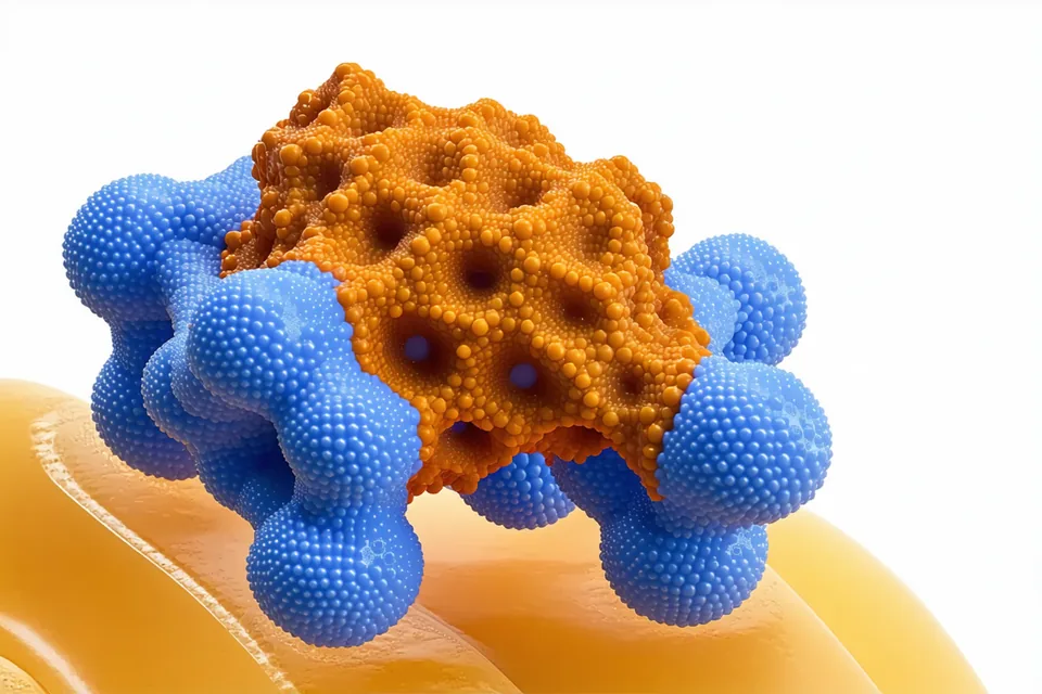
SBDD uses the three-dimensional structure of a target protein — typically from X-ray crystallography or cryo-EM — to guide compound selection and optimization. The binding site geometry constrains which molecular shapes can bind, which functional groups can make productive contacts, and which scaffold trajectories are feasible for a hit-to-lead campaign.
Fragment-based lead discovery (FBLD) extends SBDD by starting from small, rule-of-three-compliant molecules (MW ≤300 Da, cLogP ≤3, H-bond donors ≤3) that occupy sub-pockets. These fragments are elaborated systematically using structural feedback — growing vectors are identified from binding pose analysis, not from synthetic accessibility alone. The result is a hit series with explicit structural rationale at every branch point.
Docking methodology
Docking: pocket-specific calibration, not default parameters.
Molecular docking is a well-established computational method for predicting how small molecules bind to protein targets. The accuracy of docking predictions depends less on the docking algorithm than on the quality of the input structure and the appropriateness of the docking parameters for the specific pocket geometry.
Moleculepath calibrates docking protocols for each target by: (1) running cross-docking validation against any available co-crystal structures for the same target or close orthologs; (2) optimizing the binding site box and sampling parameters based on pocket volume and conformational flexibility; (3) applying protein flexibility treatment (rigid receptor, induced fit, or ensemble docking) matched to the known conformational behavior of the target class.
Pose analysis is not limited to top-score selection. Binding mode clusters are identified and the structural consistency of each cluster is evaluated before shortlisting. A high-scoring compound with an inconsistent or physically unreasonable binding pose does not make the shortlist regardless of its docking score.
FEP perturbation
Free energy perturbation: applied selectively, not by default.
Free energy perturbation (FEP) is the most accurate computational method for predicting relative binding affinities between structurally similar compounds. It is also computationally intensive — a single FEP calculation requires significant sampling and can take hours to days depending on the perturbation size and structural complexity.
Moleculepath applies FEP selectively to the top-ranked hits from an initial docking screen when structural confidence warrants it. The selection criteria for FEP escalation: the compound has a high-confidence docking pose, there are structurally analogous compounds in the hit set for perturbation, and the program team has identified specific SAR questions that FEP can answer more accurately than docking alone.
FEP is not applied as a validation stamp on all docking results. It is a targeted tool for specific binding affinity questions when the program is at a stage where the extra accuracy justifies the computational cost.
Fragment elaboration
Fragment-to-lead: growing vectors from structural analysis.
A fragment that binds with low affinity but in a well-defined binding pose is more valuable as a starting point than a high-affinity but structurally ambiguous hit from a diversity screen. Fragment elaboration begins by identifying growing vectors — atoms or positions on the fragment where additional molecular complexity can be added without disrupting the key pharmacophoric contacts.
Scaffold hopping is a parallel process: for fragments with multiple feasible elaboration vectors, the analysis identifies which molecular frameworks (not just which substitutions) can occupy the same binding sub-pocket with different pharmacokinetic properties or IP positioning. This is particularly relevant for target classes where one chemical series dominates the patent landscape — the goal is to identify a differentiated scaffold early, not to iterate on a known chemotype.
Validation benchmarks
Internal benchmark performance on published targets.
Moleculepath's docking protocols are validated on published benchmark sets. The following metrics reflect internal performance on targets with deposited crystal structures and known active sets. These benchmarks use standard virtual screening evaluation methods — results are consistent with published best-practice performance in the literature for carefully calibrated docking protocols.
| Target Class | Benchmark Set | AUC-ROC | EF 1% | Note |
|---|---|---|---|---|
| Kinases | DUD-E (18 kinase targets) | 0.82 ± 0.07 | 22–38× | Protocol calibrated per-target |
| GPCRs | GPCR-Bench (8 targets) | 0.76 ± 0.09 | 15–28× | Orthosteric site; co-crystal available |
| Proteases | DUD-E (6 protease targets) | 0.85 ± 0.05 | 30–52× | Rigid receptor; well-defined S1 pocket |
| Epigenetic | CASF-2016 (3 targets) | 0.79 ± 0.08 | 18–34× | BRD4, PCAF, EZH2 analogues |
AUC-ROC: area under the ROC curve. EF 1%: enrichment factor at 1% of the screened library. Results are internal benchmarks; published DUD-E and CASF literature ranges used for calibration. Benchmark performance does not guarantee equivalent enrichment on novel targets without validated co-crystal data.
The methodology is precise. So is the engagement.
Start with your target structure. Moleculepath returns a campaign design proposal that shows how the methodology applies to your specific binding site.
Discuss a Target